Persistent Epithelial Defects (PEDs)

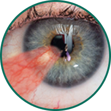
Common diagnoses resulting in or associated with corneal defects include:

5 close-up eye images depicting 5 ocular conditions that may result in or be associated with persistent epithelial defects
PEDs
5 close-up eye images depicting 5 ocular conditions that may result in or be associated with persistent epithelial defects
Superficial Punctate Keratitis
5 close-up eye images depicting 5 ocular conditions that may result in or be associated with persistent epithelial defects
Neurotrophic
 Keratitis
5 close-up eye images depicting 5 ocular conditions that may result in or be associated with persistent epithelial defects
Anterior Basement Membrane Dystrophy
5 close-up eye images depicting 5 ocular conditions that may result in or be associated with persistent epithelial defects
Pterygium

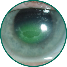
PEDs are corneal epithelial defects that fail to heal within 2 weeks and arise from diverse etiologies1

Category Etiologies of PEDs
Surgical
  • Incisional or laser-based ocular surgeries
    • Cataract surgery
    • Pterygium removal surgery
    • Laser-assisted in situ keratomileusis (LASIK)
    • Penetrating or lamellar keratoplasty
  • Neurosurgeries causing damage to the trigeminal ganglion
Injury
  • Exogenous injury
    • Chemical or thermal burns
    • Ultraviolet light injury
    • Exposure keratopathy
    • Prolonged overuse of contact lenses
  • External agents
    • Viral infection (eg, herpetic keratitis)
    • Drugs (eg, Stevens-
      Johnson
      syndrome)
Autoimmune
  • Sjögren syndrome
  • Ocular cicatricial pemphigoid
  • Ectodermal dysplasia
Allergic
  • Vernal keratoconjunctivitis
  • Atopic keratoconjunctivitis
Other
  • Vitamin A
    deficiency
  • Severe dry eye
    disease
  • Corneal
    dystrophies

PEDs share common pathophysiological drivers1:

1. Tear disorder (dry eye)


Inadequate tear film leads to reduced lubrication and excessive friction between the eyelid and the cornea, causing mechanical and inflammatory damage

2. Limbal stem cell deficiency

Disruption of the limbal stem cell niche reduces the regenerative capacity of epithelial cells

3. Inflammation


Elevated proinflammatory mediators 
(eg, IL-1β, TNF, IL-6, IL-8, matrix metalloproteinases) contribute to extracellular matrix (ECM) degradation and epithelial cell loss

4. Neurotrophic keratopathy

Alterations in corneal nerves lead to impaired sensory and trophic function, resulting in breakdown of the corneal epithelium

IL-1β, interleukin-1β; IL-6, interleukin-6; IL-8, interleukin-8; TNF, tumor necrosis factor.

Serious wounds require intervention beyond ocular surface lubrication and discontinuation of preservative-containing medications1

Human AMT-derived allografts

The gold-standard physical barrier for ocular wound repair1,2

How AMT helps heal ophthalmic wounds

  • Acts as a physical barrier, protecting the eye against mechanical trauma from lid closure and preventing surface desiccation by maintaining a hydrated microenvironment1
  • Enriched with growth factors and structural proteins that promote corneal healing2
  • Serves as a substrate for adhesion, migration, and proliferation of corneal epithelial cells and limbal stem cells when used as a graft to treat corneal pathologies1
Diagram of human amnion membrane layers (epithelium, basement membrane, compact, and fibroblast), showing the use of decellularized basement membrane in a 1 or 3-layer structure.

Only DBM offers all the benefits of AMTs, without donor cell debris, while preserving ECM functionality and triggering an efficient host cell–mediated response3,4

AMT, amniotic membrane tissue.

DBM represents an advance over cryopreserved and conventional dehydrated AMTs. The decellularization process:

Removes donor cell debris and proinflammatory chorion to minimize immune responses4
Preserves the matrix scaffold and ECM protein production4

Adheres efficiently to reduce slippage and allow for glueless, sutureless surgery5,6

References: 1. Thia ZZ, Ho YT, Shih KC, Tong L. New developments in the management of persistent corneal epithelial defects. Surv Ophthalmol. 2023;68(6):1093-1114. 2. Tehrani FD, Firouzeh A, Shabani I, Shabani A. A review on modifications of amniotic membrane for biomedical applications. Front Bioeng Biotechnol. 2021;8:1-25. 3. BIOVANCE 3L Ocular. Package insert. 4. Mao Y, Protzman NM, John N, et al. An in vitro comparison of human corneal epithelial cell activity and inflammatory response on differently designed ocular amniotic membranes and a clinical case study. J Biomed Mater Res B Appl Biomater. 2023;111(3):684-700. 5. Linsey K. Use of an eyelid pressure patch concomitantly with a decellularized dehydrated amniotic membrane for ocular surface disease management. Ophthalmol Ther. 2025;14(3):573-584. 6. Rivera-Morales P, Barnard L, Linderman W, Gill M, Diaz V. Surgical time and postoperative symptoms study in pterygium excision and amniotic membrane graft using Celularity triple layer dehydrated amniotic membrane. Clin Ophthalmol. 2023;17:1967-1974.

Thank you!

Keep an eye out for emails with the latest news from DefEYE, Inc.

Add Your Heading Text Here

Skip to content
DefEYE™, Inc. Logo
Privacy Overview

This website uses cookies so that we can provide you with the best user experience possible. Cookie information is stored in your browser and performs functions such as recognising you when you return to our website and helping our team to understand which sections of the website you find most interesting and useful.